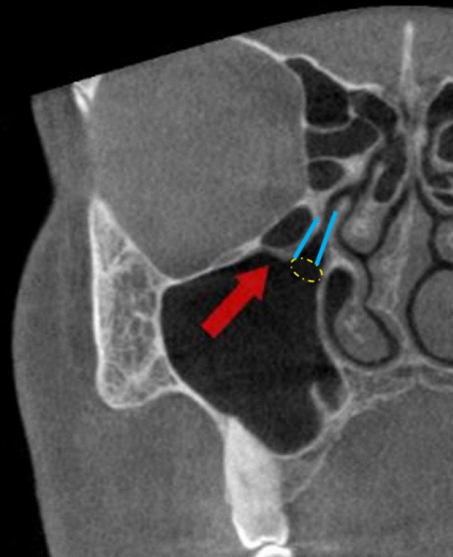
Haller Cells Radiology - Frontal Recess Air Cells Spectrum Of Ct Appearances Coates 2003 Australasian Radiology Wiley Online Library / Haller's cells, also known as infraorbital ethmoid cells are located at the medial roof of the maxillary sinus in the inferior most portion of the lamina papyracea.
Haller Cells Radiology - Frontal Recess Air Cells Spectrum Of Ct Appearances Coates 2003 Australasian Radiology Wiley Online Library / Haller's cells, also known as infraorbital ethmoid cells are located at the medial roof of the maxillary sinus in the inferior most portion of the lamina papyracea.. In the ct image haller cells are seen. The indian journal of radiology & imaging. Infraorbital ethmoid (haller) cells are extensions of the anterior ethmoid sinus into the floor of the orbit and superior aspect of the maxillary sinus. Haller's cells, also known as infraorbital ethmoid cells are located at the medial roof of the maxillary sinus in the inferior most portion of the lamina papyracea. There is an intraoperative relationship of the left haller cell to the maxillary sinus.
In the ct image haller cells are seen. The cells are variable in both size and number in the lateral mass of each of the ethmoid bones and cannot be palpated during an extraoral examination. Concha bullosa and haller cell resection, maxillary sinus outflow obstruction. Haller cells lay posterosuperior to the natural maxillary os. Named after albrecht von haller, a swiss anatomist.
Jaypeedigital Ebook Reader from d45jl3w9libvn.cloudfront.net Dendritic cells (dcs) and monocytes play a central role in pathogen sensing, phagocytosis, and antigen presentation and consist of multiple specialized subtypes. There is an intraoperative relationship of the left haller cell to the maxillary sinus. Բջջային հեռախոսների, պլանշետների և աքսեսուարների մասնագիտացված խանութ սրահ They are extramuralethmoidal air cells that extend into the inferomedial orbitalfloor and are present in ~20. In the ct image haller cells are seen. It may narrow the ipsilateral ostiomeatal. Performance of radiologists in differentiating covid 19 from viral pneumonia on chest ct radiology. Variation of posterior ethmoid cells located above the sphenoid sinus as a result of hyperpneumatization.
May be due to allergy, a viral or bacterial infection or.
The aim of this retrospective study was to evaluate the. Concha bullosa and haller cell resection, maxillary sinus outflow obstruction. Partial opacification of the bilateral mastoid air cells, middle ear, ethmoid air cells. May be due to allergy, a viral or bacterial infection or. Haller's cells are anatomical variants of the paranasal sinuses, which were first described by albrecht von haller in 1765. View of lateral nasal wall (turbinates removed). Haller cells | radiology reference article. Haller cells are one of the anatomical variations in the orbital area, which are important in endoscopic surgical procedures and have a role in the pathogenesis of some diseases. Radial glial cells (rgcs) are a morphologically, biochemically, and functionally distinct nonneuronal cell class that, during development, radially spans the entire width of the developing cerebral wall. There is an intraoperative relationship of the left haller cell to the maxillary sinus. Learn vocabulary, terms and more with flashcards, games and other study tools. Haller cells are also known as infraorbital ethmoidal air cells or maxilloethmoidal cells. Variation of posterior ethmoid cells located above the sphenoid sinus as a result of hyperpneumatization.
The pectus or haller index is calculated by dividing the maximum transverse diameter of the chest by the ap diameter.29 the pectus index was. Imaging of sinonasal inflammatory disease. 100% aneuploidy in 1385 cells examined. Haller cells, also known as infraorbital ethmoidal air cells, are ethmoid air cells located lateral to the terminology the sphenoethmoidal air cel. See more ideas about radiology, radiography, medical.
Paranasal Sinuses Nasal Cavity And Face Radiology Key from i1.wp.com This is formed by lateral and posterior pneumatization of the most posterior ethmoid cells over the sphenoid sinus. Haller cells | radiology reference article. There is a close relation with the optic nerve. Imaging of sinonasal inflammatory disease. Haller cells, also known as infraorbital ethmoidal air cells, are ethmoid air cells located lateral to the terminology the sphenoethmoidal air cel. Variation of posterior ethmoid cells located above the sphenoid sinus as a result of hyperpneumatization. Haller cells are also known as infraorbital ethmoidal air cells or maxilloethmoidal cells. 65,098 likes · 1,328 talking about this · 225 were here.
Coronal scan of ostiomeatal unit.
Learn vocabulary, terms and more with flashcards, games and other study tools. They can obstruct the outflow tract of the maxillary sinus and must be removed when there is pathology within. There is an intraoperative relationship of the left haller cell to the maxillary sinus. The cells are variable in both size and number in the lateral mass of each of the ethmoid bones and cannot be palpated during an extraoral examination. See more ideas about radiology, radiography, medical. Concha bullosa and haller cell resection, maxillary sinus outflow obstruction. In the ct image haller cells are seen. Haller cells, also known as infraorbital ethmoidal air cells, are ethmoid air cells located lateral to the terminology the sphenoethmoidal air cel. Haller cells lay posterosuperior to the natural maxillary os. View of lateral nasal wall (turbinates removed). Imaging of sinonasal inflammatory disease. Four typical hela marker chromosomes have been reported in the literature. Radial glial cells (rgcs) are a morphologically, biochemically, and functionally distinct nonneuronal cell class that, during development, radially spans the entire width of the developing cerebral wall.
In the ct image haller cells are seen. See more ideas about radiology, radiography, medical. In the ct image haller cells are seen. At first an access to the. Haller cells, also known as infraorbital ethmoidal air cells, are ethmoid air cells located lateral to the terminology the sphenoethmoidal air cel.
Cbct Analysis Of Haller Cells Relationship With Accessory Maxillary Ostium And Maxillary Sinus Pathologies Springerlink from media.springernature.com Imaging of sinonasal inflammatory disease. There is a close relation with the optic nerve. There is an intraoperative relationship of the left haller cell to the maxillary sinus. Haller cells, also known as infraorbital ethmoidal air cells, are ethmoid air cells located lateral to the terminology the sphenoethmoidal air cel. The pectus or haller index is calculated by dividing the maximum transverse diameter of the chest by the ap diameter.29 the pectus index was. Haller cells are anterior ethmoid air cells extending into the maxillary sinus (figure 17). Haller's cells, also known as infraorbital ethmoid cells are located at the medial roof of the maxillary sinus in the inferior most portion of the lamina papyracea. View of lateral nasal wall (turbinates removed).
Haller cells are also known as infraorbital ethmoidal air cells or maxilloethmoidal cells.
Performance of radiologists in differentiating covid 19 from viral pneumonia on chest ct radiology. Haller's cells, also known as infraorbital ethmoid cells are located at the medial roof of the maxillary sinus in the inferior most portion of the lamina papyracea. Haller cells, also known as infraorbital ethmoidal air cells, are ethmoid air cells located lateral to the terminology the sphenoethmoidal air cel. Epidemiology they are present in ~20. Haller cells lay posterosuperior to the natural maxillary os. Haller's cells are anatomical variants of the paranasal sinuses, which were first described by albrecht von haller in 1765. Learn vocabulary, terms and more with flashcards, games and other study tools. Top 3 differentials in radiology: View of lateral nasal wall (turbinates removed). In the ct image haller cells are seen. This is formed by lateral and posterior pneumatization of the most posterior ethmoid cells over the sphenoid sinus. The pectus or haller index is calculated by dividing the maximum transverse diameter of the chest by the ap diameter.29 the pectus index was. The ethmoid sinuses or ethmoid air cells of the ethmoid bone are one of the four paired paranasal sinuses.